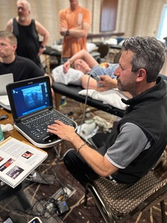
Search
Close this search box.

Musculoskeletal (MSK) Ultrasound Home Study Courses

Professions that benefit from MSK Ultrasound

Physical Therapist, Chiropractors, Athletic Trainers,  Occupational Therapists,  Physicians, Nurse Practitioners, and Physician Assistants,  

COURSE INFORMATION

Dr. Randy Moore
Description

Diagnostic ultrasound is quickly becoming the standard for musculoskeletal (MSK) care. MSK US is not a new tool but manual therapists, orthopedic practitioners and other rehab professionals are now, more than ever, understanding its clinical value. Adding MSK US to your evaluation improves the validity and reliability of your MSK diagnosis by viewing real-time images and motion analysis. Being able to visualize the soft tissue allows the clinician the ability to not only deliver the most effective treatment but to track soft tissue healing in a way that has not been readily available before. The MSK US home study videos provide a solid understanding of MSK imaging allowing recognition of normal and abnormal anatomy that are at the root of the commonly encountered injuries seen in clinical practice.


This course helps you create a foundation in the emerging field of MSK Ultrasound. You’ll learn musculoskeletal anatomy and sonoanatomy, probe placement, ultrasound guided imaging fundamentals and practical examples, pathology and ultrasound guided injections. Get in-depth training today and gain a solid understanding of the musculoskeletal ultrasound field, with plenty of virtual ‘hands-on’ learning that enables you to truly become an MSK Master.

Prerequisite

No prior diagnostic ultrasound or dry needling training or experience is required. 

Learn about the Instructor

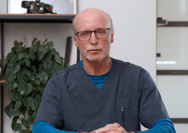
Dr. Moore is an extremely highly regarded and sought after pioneer, teacher and author in the quickly emerging field of musculoskeletal sonography. He teaches using straightforward and easy to understand teaching methods that have been deemed essential to shortening the learning curve for medical practitioners, medical sonographers, physician assistants, nurse practitioners, physical therapists and allied health professionals as they develop expertise in MSK sonography.

>> LEARN MORE

LOWER EXTREMITY INFO
LEARNING OBJECTIVES:
  1. Establish a solid understanding of MSK ultrasound imaging fundamentals
  2. Recognize normal ultrasound anatomy of the Lower Extremity (hip, knee, foot & ankle and lumbar spine)
  3. Be able to recognize abnormal ultrasound anatomy of the Lower Extremity
  4. Understand common Lower Extremity pathology


AGENDA:
 
Module 1: Introduction
  • This module provides an Introduction to basic Grayscale Imaging and Normal Sonoanatomy
Module 2: Imaging Fundamentals
  • Session 1: Reviews the crucial nature of knowing normal sonoanatomy.
  • Session 2: Reviews Image Acquisition, Image Orientation and Image Interpretation Algorithm.
  • Session 3: Reviews normal Sonoanatomy. Clearly understanding normal sonoanatomy precedes accurate identification of pathology.
Module 3: The Hip
  • Overview: The Hip – More than Bursitis
  • Session 1: presents the single Anterior Hip image which yields much diagnostic information
  • Session 2: presents the Ilio-Psoas Tendon, Greater Trochanteric Tendons (GMin and GMed)
  • Session 3: the Piriformis Muscle, Sciatic Nerve, Prosthetic Hips, Lateral Femoral Cutaneous Nerve
Module 4: The Knee
  • Overview: The Knee – Above, Below and All Around
  • Session 1: presents the Supra-Patellar Recess which is a common site for guided injections, Femoral Trochlea to evaluate osteoarthritis
  • Session 2: teaches the Infra-PatellarTendon, Hoffa’s Fat Pad, Lateral Collateral Ligament (LCL), Peroneal Nerve
  • Session 3: the Medial Collateral Ligament (MCL), Medial Meniscus,Pes Anserine Bursa, Medial Plica Syndrome, Medial Patello Femoral Ligament (MPFL) involved in Patellar tracking diagnosis
  • Session 4: the Popliteal Fossa, Baker’s Cyst, Popliteal Neuro-Vascular Bundle
Module 5: The Foot & Ankle
  • Overview: The Foot & Ankle: Achilles, Plantar Fascia and More
  • Session 1: presents the ExtensorTendons of the toes, Tib-Talar Joint (Ankle Joint), Deep Peroneal Nerve, Anterior Talo-Fibular (ATFL) Ligament, Tib-Fib Ligament (High Ankle Sprains)
  • Session 2: presents the Peroneal Tendons, Calcaneo-Fibular (CFL) ligament
  • Session 3: presents the Medial Ankle (Tarsal Tunnel), Deltoid Ligaments
  • Session 4: presents the Achilles Tendon, Saphenous Vein, Sural Nerve, Medial Sub-Talar Joint
  • Session 5: presents Plantar Fascia, Plantar Plate, 1st Metatarso-Phalangeal Joint (MTP), Flexor Hallucis Longus (FHL) Tendon, Inter-digital lesions
Module 6: The Lumbar Spine
  • Overview: The Spine – Lumbar Spine
  • Session 1: presents the transducer selection for spinal imaging, patient position, the “Navigation Triangle”, the two long axis(LAX) angles of insonation, the Erector Spinae Muscles and Multifidus Muscles
  • Session 2: presents the Short Axis Imaging, Facet Joints, the “King Sign” and Facet Joint innervation
  • Session 3: presents the Sacro-Iliac Joint, SI Joint Injection technique, Long Posterior SI Ligaments, Ilio-Lumbar Ligament, Caudal Epidural Space
Module 7: Lower Extremity Pathology
  • Overview: Common lower extremity pathology
  • Session 1: presents the Anterior Hip Capsule, Acetabular Labrum, Psoas Tendon, Gluteal Tendon (GMin,GMax) insertions on the Greater Trochanter, Snapping Hip Syndromes
  • Session 2: presents the Sciatic Nerve and Piriformis Muscle imaging, Hamstring Tendons (Semi-Membranosus, Conjoined Tendons)
  • Session 3: presents the Suprapatellar Effusions, Quadriceps Tendon, Femoral Trochlea Osteoarthritis, Bi-Partite Patella, Infra-Patellar Tendinosis, Osgood Schlatter’s Disease, Infra-Patellar Fat Pad Impingement
  • Session 4: presents the Medial Menicus Tears, Medial Collateral Ligament (MCL) Tears, Pes Anserine Bursa, Medial Plica Syndrome
  • Session 5: presents the Ilio-Tibial Band (ITB), the often neglected Tibio-Fibular Joint, Lateral Collateral Ligament (LCL), Peroneal Nerve, Popliteal Fossa and Baker’s Cyst, Tibial Neuro-Vascular Bundle
  • Session 6: the the Tibio-Talar Joint Effusions, Anterior Talo-Fibular (ATFL), Calcaneo-Fibular (CFL) Ligaments, Tib-Fib Ligament (High Ankle Sprain), Lisfranc Injury (Mid-Foot Disruption), Tibialis Posterior Tendon Rupture, Plantar Foot Lipoma, Plantar Fasciitis, Inter-Digital Neuroma, Plantar Plate Fracture
Module 8: Ultrasound Guided Injections
  • Overview: presents the needed mechanics and ergonomics to perform accurate ultrasound guided injections which produced optimal outcomes.
  • Session 1: discusses ultrasound images and the importance it plays in injections
  • Session 2: discusses Maintaining Sterile Field, Optimal Ergonomics for the procedure, Initial Needle Advancement (Puncture Site) relative to depth of target, suggestions for Needle Localization, and maintaining “Perpendicularity” to the ultrasound beam.
  • Session 3: discusses Out Of Plane Injections, The “Falling Star”, Beam Steering to enhance needle perpendicularity, Guided Injection Examples: Sub-Acromial/Deltoid, Gleno-Humeral Joint,Median Nerve, DeQuervain’s Out Of Plane, Medial Collateral Ligament, Calcific Tendinosis Supraspinatus, Baker’s Cyst Aspiration, Lateral Epicondyle, Intra-Muscular, Plantar Foot/Heel.
UPPER EXTREMITY INFO
LEARNING OBJECTIVES:
  1. Established a solid understanding of MSK ultrasound imaging fundamentals
  2. Recognize normal ultrasound anatomy of the Upper Extremity (shoulder, elbow and hand/wrist
  3. Be able to recognize abnormal ultrasound anatomy of the Upper Extremity
  4. Understand common upper extremity pathology


AGENDA:

Module 1: Introduction

  • This module provides an Introduction to basic Grayscale Imaging and Normal Sonoanatomy
Module 2: Imaging Fundamentals
  • Session 1: Reviews the crucial nature of knowing normal sonoanatomy.
  • Session 2: Reviews Image Acquisition, Image Orientation and Image Interpretation Algorithm.
  • Session 3: Reviews normal Sonoanatomy. Clearly understanding normal sonoanatomy precedes accurate identification of pathology.
Module 3: The Shoulder
  • Overview: The Shoulder – The Most Mobile & Vulnerable Joint
  • Session 1: presents Biceps Tendon at the bicipital groove and distally near the Pec Major Tendon
  • Session 2: presents the Supraspinatus Tendon and Rotator Cuff Interval anatomy and function
  • Session 3: presents the final two of the four Rotator Cuff Tendons… Infraspinatus Tendon and Teres Minor Tendon
  • Session 4: presents Acromio-Clavicular Joint imaging and Anterior Shoulder Impingement, then discusses posterior shoulder images of the Gleno-Humeral Joint, Spino-Glenoid Notch.
Module 4: The Elbow
  • Overview: The Elbow – A 360 Degree Evaluation
  • Session 1: presents the cross-sectional anatomy of the Anterior Elbow, Coronoid Fossa, Distal Biceps Tendon at the Radial insertion.
  • Session 2: teaches the Antero-Lateral Elbow Joint, Lateral Epicondyle(Common Extensor Insertion) and Medial Epicondyle (Common Flexor Insertion)
  • Session 3: presents Posterior Elbow and Olecranon Fossa, Triceps Tendon/Muscle, Ulnar Nerve imaging at the Ulnar Groove
Module 5: The Hand & Wrist
  • Overview: The Hand & Wrist – Complex Anatomy
  • Session 1: presents the Median Nerve Localization, Carpal Tunnel Measurement, Ulnar Nerve at Guyon’s Canal, Duyputren’s Contracture.
  • Session 2: presents the Dorsal Wrist Compartments 1 thru 3, DeQuervain’s Tenosynovitis, Dorsal MCP Joint
  • Session 3: presents A1-A2 Pulley/Trigger Finger, 1st CMC (Carpal Meta-Carpal) Joint, Ulnar Collateral Ligament.
  • Session 4: presents the Triangular Fibrocatilage Complex (TFCC)
Module 6: Upper Extremity Pathology
  • Overview: Common upper extremity pathology
  • Session 1: presents the most common tendon pathology… Tendinosis.
  • Session 2: presents the Subscapularis Tears, Sub-Deltoid Bursa, Sub-Cutaneous Lipoma, Biceps Tenosynovitis (Fluid inside the Tendon Sheath), Subluxed/Dislocated Biceps.
  • Session 3: presents the Olecranon Synovitis of the Posterior Elbow, Subluxing/Dislocating Ulnar Nerve, Lateral Epicondyle.
  • Session 4: presents the Dorsal Ganglion Cyst, DeQuervain’s StenosingTenosynovitis, DePuytren’s Contracture, Median Nerve/Carpal Tunnel Syndrome, Snapping Extensor Carpi Ulnaris Tendon, Dorsal Meta-Carpal Phalangeal (MCP)Joint, Synovial Fluid vs Synovial Hypertrophy.
Module 7: Ultrasound Guided Injections
  • Overview: presents the needed mechanics and ergonomics to perform accurate ultrasound guided injections which produced optimal outcomes.
  • Session 1: discusses ultrasound images and the importance it plays in injections
  • Session 2: discusses Maintaining Sterile Field, Optimal Ergonomics for the procedure, Initial Needle Advancement (Puncture Site) relative to depth of target, suggestions for Needle Localization, and maintaining “Perpendicularity” to the ultrasound beam.
  • Session 3: discusses Out Of Plane Injections, The “Falling Star”, Beam Steering to enhance needle perpendicularity, Guided Injection Examples: Sub-Acromial/Deltoid, Gleno-Humeral Joint,Median Nerve, DeQuervain’s Out Of Plane, Medial Collateral Ligament, Calcific Tendinosis Supraspinatus, Baker’s Cyst Aspiration, Lateral Epicondyle, Intra-Muscular, Plantar Foot/Heel.

MSK Home Study Courses are approved for CEUs for Chiropractors and Athletic Trainers!

PURCHASE OPTIONS

LEARN MSK ULTRASOUND TODAY!

Get both at a special discounted price!

LOWER EXTREMITY COURSE

This on-demand video series will establish a solid understanding of MSK ultrasound imaging fundamentals. Emphasis is on recognizing normal and abnormal ultrasound anatomy of the hip, knee, ankle, and foot. The lumbar spine will also be discussed!

$550
  • Understand the common lower extremity pathologies seen in clinical practice
  • Understand the ins and outs of musculoskeletal ultrasound scanners
  • Understand the role of musculoskeletal ultrasound in clinical practice
  • Understand the mechanics and ergonomics needed to perform accurate ultrasound guided injections & dry needling to produce optimal outcomes
  • Access to our MSK ultrasound report templates

MSK ULTRASOUND BUNDLE

Get both courses together and save! Want to improve your assessment and treatment skills for upper and lower extremity conditions?  Start on your journey toward the future of MSK clinical care!

$995 regularly $1,100
  • Understand the common upper AND lower extremity pathologies seen in clinical practice
  • Understand the ins and outs of musculoskeletal ultrasound scanners Understand the role of musculoskeletal ultrasound in clinical practice
  • Understand the ins and outs of musculoskeletal ultrasound scanners
  • Understand the mechanics and ergonomics needed to perform accurate ultrasound guided injections and dry needling to produce optimal outcomes
  • Access to our MSK ultrasound report templates

UPPER EXTREMITY COURSE

This on-demand video series will establish a solid understanding of MSK ultrasound imaging fundamentals. Emphasis is on recognizing normal and abnormal ultrasound anatomy of the shoulder, elbow, wrist and hand.

$550
  • Understand the common upper extremity pathologies seen in clinical practice
  • Understand the ins and outs of musculoskeletal ultrasound scanners
  • Understand the role of musculoskeletal ultrasound in clinical practice
  • Understand the mechanics and ergonomics needed to perform accurate ultrasound guided injections and dry needling to produce optimal outcomes
  • Access to our MSK ultrasound report templates

QUESTIONS ABOUT MSK ULTRASOUND?

FLASH SALE!

20% OFF

any IDN Course!

*This discount is valid for new registrations only and can not be combined with other discount codes.  Offer Expires: 12/31/2023

Integrative Dry Needling Logo Orange

Not sure which course is right for you? No problem – we created an intuitive process to help!